Thursday, October 27, 2005

The stunning beauty of Cell Biology - man's advance at the hand of Art
microbe-1


microbe-2


microbe-3


microbe-4


microbe-5


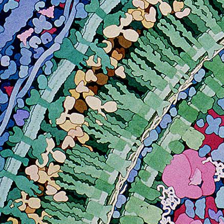
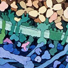
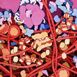
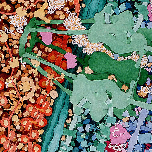
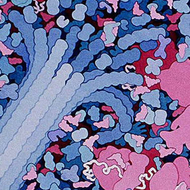
These amazing images are to-scale watercolor illustrations of a bacterium and blood cells. These are small sections of a larger piece made by Dr. David Goodsell. Notes on this work say

Macrophages circulate through the blood, searching for bacterial infection. When bacteria are found, macrophages engulf and digest them. This series of three paintings shows a macrophage engulfing a bacterium. Only a portion of the two cells, where a pseudopod of the macrophage is extending over the bacterium, is shown. The original paintings are 1 meter tall--at this magnification, the macrophage would fill most of a building.


David S. Goodsell is someone I admire deeply for his ability to illustrate Cell Biology (my favorite part of the life sciences, cant help the bias).

He is an associate professor in the Department of Molecular Biology at the Scripps Research Institute in La Jolla, California. He is the author of Bionanotechnology: Lessons from Nature (J. Wiley and Sons, 2004), Our Molecular Nature: The Body's Motors, Machines, and Messages (Springer-Verlag, 1996), and The Machinery of Life (Springer-Verlag, 1993). His recent biomolecular artwork can be viewed here. Address: Department of Molecular Biology, The Scripps Research Institute, La Jolla, CA 92037. Internet: goodsell@scripps.edu (I got this blurb from here)

Read about his scientific interests here.

Read about his artistic work here.

A nice online presentation he has on Overview of Biological Machines from the Designing Nanostructures: A tutorial site can be found here. And you can pull up a nice slide presentation here.

Here is a lovely poster on Molecular Machinery (PDF 5MB)

Wednesday, October 12, 2005

Computational model of dynein-dependent self-organization of microtubule asters

Computational model of dynein-dependent self-organization of microtubule asters (PDF)
E. N. Cytrynbaum, V. Rodionov and A. Mogilner Journal of Cell Science 117, 1381-1397 (2004)

Polar arrays of microtubules play many important roles in the cell. Normally, such arrays are organized by a centrosome anchoring the minus ends of the microtubules, while the plus ends extend to the cell periphery. However, ensembles of molecular motors and microtubules also demonstrate the ability to self-organize into polar arrays. We use quantitative modeling to analyze the self-organization of microtubule asters and the aggregation of motor-driven pigment granules in fragments of fish melanophore cells. The model is based on the observation that microtubules are immobile and treadmilling, and on the experimental evidence that cytoplasmic dynein motors associated with granules have the ability to nucleate MTs and attenuate their minus-end dynamics. The model explains the observed sequence of events as follows. Initially, pigment granules driven by cytoplasmic dynein motors aggregate to local clusters of microtubule minus ends. The pigment aggregates then nucleate microtubules with plus ends growing toward the fragment boundary, while the minus ends stay transiently in the aggregates. Microtubules emerging from one aggregate compete with any aggregates they encounter leading to the gradual formation of a single aggregate. Simultaneously, a positive feedback mechanism drives the formation of a single MT aster – a single loose aggregate leads to focused MT nucleation and hence a tighter aggregate which stabilizes MT minus ends more effectively leading to aster formation. We translate the model assumptions based on experimental measurements into mathematical equations. The model analysis and computer simulations successfully reproduce the observed pathways of pigment aggregation and microtubule aster self-organization. We test the model predictions by observing the self-organization in fragments of various sizes and in bi-lobed fragments. The model provides stringent constraints on rates and concentrations describing microtubule and motor dynamics, and sheds light on the role of polymer dynamics and polymer-motor interactions in cytoskeletal organization.

Key words: Cytoskeleton, Self-organization, Mathematical model, Molecular motors, Microtubules, Mitosis


Fig. 5. 1D implementation of the model in a long narrow fragment of length 2L. Two dynamic sub-populations of MTs with opposite orientations are characterized by the densities of the plus (pr,l) and minus (mr,l) ends and the polymer densities Nr,l. Three pigment sub-populations are described by the densities of the granules gliding to the right (gr) and left (gl) with speed vg, and static granules (gs) dissociated from the MTs.


Fig. 6. Illustration of the numerical implementation of the 2D computational model. Each MT generates an effective velocity field (arrows) in the rectangular domain of influence (see isolated MTs at the periphery). The corresponding velocities are minus-end-directed and decrease away from the MT. The velocity fields of individual MTs add locally and geometrically. Note that the velocity field in the center is not parallel to any individual MT but results from the vector sum of individual contributions. The shading shows the granule density generated fast by the shown effective velocity field.


Fig. 8. Computer simulations of the 2D model (compare with Figs 1 and 2). MTs are shown as segments. The granule density is illustrated by shading with darker areas corresponding to higher densities. (A) Initially, the MTs are distributed randomly and the granule density is uniform. (B) The granules quickly aggregate into a few local foci before MTs can re-organize. (C) The local aggregates coalesce into a single loose aggregate over a few minutes simultaneously with the re-organization of the MTs. (D) The pigment aggregate tightens over the next few minutes and the MT aster is clearly seen.


Fig. 9. MTs and granule density are illustrated as in Fig. 8. Level-curves of granule density are added for emphasis. Both images show the state of a fragment soon after dynein stimulation. (A) At low and moderate nucleation rates, a few local aggregates evolve initially. (B) At high nucleation rates, the granules initially coalesce into a single loose aggregate.


Fig. 10. Self-organization in the bi-lobed fragment. (Left) Phase contrast images show pigment distribution. (Right) Results of a computer simulation in the bi-lobed fragment. The experimental images are obtained before the adrenalin treatment, and 5 and 10 minutes after it, respectively. Bars, 10 µm.

Cell Cycle computational models

Found this interesting resource on computational models of the cell cycle (PDF doc)

Development and validation of computational models of cellular interaction
R H Smallwood, W M L Holcombe, D C Walker (2004)

----

BioInformatics Reviewed Web page Links - a nice compendium of intro bioinformatics tutorials and learning opportunities.


-----

Tuesday, September 13, 2005

Gene expression and in vitro development of inter-species nuclear transfer embryos

Gene expression and in vitro development of inter-species nuclear transfer embryos
Mol. Reprod. Dev. 66: 334-342, 2003

Sezen Arat, S. Jacek Rzucidlo, Steven L. Stice

Abstract
This study examined the chromatin morphology, in vitro development, and expression of selected genes in cloned embryos produced by transfer of mouse embryonic fibroblasts (MEF) into the bovine ooplasm. After 6 hr of activation, inter-species nuclear transfer (NT) embryos (MEF-NT) had one (70%) or two pronuclei (20%), respectively. After 72 hr of culture in vitro, 62.6% of the MEF-NTs were arrested at the 8-cell stage, 31.2% reached the 2- to 4-cell stage, and only 6.2% had more than eight blastomeres, but none of these developed to the blastocyst stage. Whereas, 20% of NT embryos derived from bovine embryonic fibroblast fused with bovine ooplasm (BEF-NT) reached the blastocyst stage. Donor MEF nuclei expressing an Enhanced Green Fluorescent Protein (EGFP) transgene resulted in 1- to 8-cell stage MEF-NT that expressed EGFP. The expression of selected genes was examined in 8-cell MEF-NTs, 8-cell mouse embryos, enucleated bovine oocytes, and MEFs using RT-PCR. The mRNA for heat shock protein 70.1 (Hsp 70.1) gene was detected in MEF-NTs and MEF, but not in mouse embryos. The hydroxy-phosphoribosyl transferase (HPRT) mRNA was found in normal mouse embryos and MEF but not in MEF-NTs. Expression of Oct-4 and embryonic alkaline phospatase (eAP) genes was only detected in normal mouse embryos and not in the inter-species NT embryos. Abnormal gene expression profiles were associated with an arrest in the development at the 8-cell stage, but MEF-NT embryos appeared to have progressed through gross chromatin remodeling, typical of intra-species NT embryos. Therefore, molecular reprogramming rather than chromatin remodeling may be a better indicator of nuclear reprogramming in inter-species NT embryos. Mol. Reprod. Dev. 66: 334-342, 2003. © 2003 Wiley-Liss, Inc.

link for abstract

NT: Gene Expression Patterns

Gene Expression Patterns in Bovine In vitro-Produced and Nuclear Transfer-Derived Embryos and Their Implications for Early Development "Cloning and Stem cells" Mar 2002, Vol. 4, No. 1: 29-38

H. Niemann, C. Wrenzycki, A. Lucas-Hahn, T. Brambrink, W.A. Kues, J.W. Carnwath

Bovine in vitro-produced (IVP) and nuclear transfer (NT)-derived embryos differ from their in vivo-developed counterparts in a number of characteristics. A preeminent observation is the occurrence of the large offspring syndrome, which is correlated with considerable embryonic fetal and postnatal losses. We summarize here results from our studies in which we compared gene expression patterns from IVP and NT-derived embryos with those from their IVP counterparts. Numerous aberrations were found in IVP and NT-derived embryos, including a complete lack of expression, an induced expression, or a significant up- or downregulation of a specific gene. These alterations may affect a number of physiological functions and are considered as a kind of stress response of the embryos to deficient environmental conditions. We hypothesize that the alterations are caused by epigenetic modifications, primarily by changes in the methylation patterns. Unravelling these epigenetic modifications is promising to reveal the underlying mechanisms of the large offspring syndrome.
This paper was cited by:
Duration of In Vitro Maturation of Recipient Oocytes Affects Blastocyst Development of Cloned Porcine Embryos
Michael Hölker, Björn Petersen, Petra Hassel, Wilfried A. Kues, Erika Lemme, Andrea Lucas-Hahn, Heiner Niemann
Cloning and Stem Cells. Mar 2005, Vol. 7, No. 1: 35-44
Abstract | Full Text PDF: For printing or With links | Related
Chromosomal instability in the cattle clones derived by somatic cell nuclear-transfer
Hirofumi Hanada, Kumiko Takeda, Takahiro Tagami, Keijiro Nirasawa, Satoshi Akagi, Noritaka Adachi, Seiya Takahashi, Yoshitaka Izaike, Masaki Iwamoto, Dai-Ichiro Fuchimoto
Molecular Reproduction and Development. 2005, Vol. 71, No. 1: 36
CrossRef
Chronological Appearance of Apoptosis in Bovine Embryos Reconstructed by Somatic Cell Nuclear Transfer from Quiescent Granulosa Cells
JO Gjorret, J Wengle, P Maddox-Hyttel, WA King
Reproduction in Domestic Animals. 2005, Vol. 40, No. 3: 210
CrossRef
Global gene expression analysis comparing bovine blastocysts flushed on day 7 or produced in vitro
M. Mohan, A.G. Hurst, J.R. Malayer
Molecular Reproduction and Development. 2004, Vol. 68, No. 3: 288
CrossRef
Animal Cloning: Reprogramming the Donor Genome
Seiya Takahashi
Journal of Mammalian Ova Research. 2004, Vol. 21, No. 3: 74
CrossRef
Cryopreservation of Bovine Somatic Cell Nuclear-Transferred Blastocysts: Effect of Developmental Stage
Dasari AMARNATH, Yoko KATO, Yukio TSUNODA
Journal of Reproduction and Development. 2004, Vol. 50, No. 5: 593
CrossRef
Developmental potential of bovine nuclear transfer embryos and postnatal survival rate of cloned calves produced by two different timings of fusion and activation
Satoshi Akagi, Noritaka Adachi, Kazutsugu Matsukawa, Masanori Kubo, Seiya Takahashi
Molecular Reproduction and Development. 2003, Vol. 66, No. 3: 264
CrossRef
Gene expression and in vitro development of inter-species nuclear transfer embryos
Sezen Arat, S. Jacek Rzucidlo, Steven L. Stice
Molecular Reproduction and Development. 2003, Vol. 66, No. 4: 334
CrossRef

NT: Abnormal gene expression

Abnormal gene expression in cloned mice derived from embryonic stem cell and cumulus cell nuclei
David Humpherys*,dagger , Kevin Eggan*,dagger , Hidenori AkutsuDagger , Adam Friedman*, Konrad Hochedlinger*, Ryuzo YanagimachiDagger , Eric S. Lander*,dagger , Todd R. Golub*,§,¶, and Rudolf Jaenisch*,dagger ,||

* Whitehead Institute for Biomedical Research and dagger Department of Biology, Massachusetts Institute of Technology, 9 Cambridge Center, Cambridge, MA 02142; Dagger Department of Anatomy and Reproductive Biology, John A. Burns School of Medicine, University of Hawaii, Honolulu, HI 96822; § Department of Pediatric Oncology, Dana-Farber Cancer Institute, Boston, MA 02115; and ¶ Department of Pediatrics, Harvard Medical School, Boston, MA 02115

link

Abstract

To assess the extent of abnormal gene expression in clones, we assessed global gene expression by microarray analysis on RNA from the placentas and livers of neonatal cloned mice derived by nuclear transfer (NT) from both cultured embryonic stem cells and freshly isolated cumulus cells. Direct comparison of gene expression profiles of more than 10,000 genes showed that for both donor cell types approx 4% of the expressed genes in the NT placentas differed dramatically in expression levels from those in controls and that the majority of abnormally expressed genes were common to both types of clones. Importantly, however, the expression of a smaller set of genes differed between the embryonic stem cell- and cumulus cell-derived clones. The livers of the cloned mice also showed abnormal gene expression, although to a lesser extent, and with a different set of affected genes, than seen in the placentas. Our results demonstrate frequent abnormal gene expression in clones, in which most expression abnormalities appear common to the NT procedure whereas others appear to reflect the particular donor nucleus.


Fig. 2. (below) Northern analysis of several genes dysregulated in NT placentas at term. Lanes 1-6 contain RNA from naturally derived B6/129 controls, whereas the RNAs in lanes 7 and 8 are derived from the placentas from normal B6/129 zygotes that had been cultured in vitro before transfer to a surrogate mother. RNAs in lanes 9-18 are from cumulus cell NT placentas of the indicated genetic backgrounds: lanes 9-13, DBA/Cast; lanes 14 and 15, 129/Cast; lane 16, AJ/Cast, and lanes 17 and 18, B6/DBA. RNAs in lanes 19-37 are from placentas of ES cell NT mice: lanes 19-29 are derived from the V6.5 (B6/129) line, lanes 30-33 are from targeted subclones (14) of the V6.5 line (lanes 30-32, subclone 89; lane 33, subclone 23), lane 34 is from the V17.2 (BALB/129) line, and lanes 35-37 are from the F1-2.3 (129/Cast) line.

Mechanistic Model for MT organization

paper link

A Mechanistic Model for the Organization of Microtubule Asters by Motor and Non-Motor Proteins in a Mammalian Mitotic Extract
Arijit Chakravarty, Louisa Howard, and Duane A. Compton *

Department of Biochemistry, Dartmouth Medical School, Hanover, New Hampshire 03755; and Rippel Electron Microscope Facility, Dartmouth College, Hanover, New Hampshire 03755

Abstract

We used computer simulation to understand the functional relationships between motor (dynein, HSET, and Eg5) and non-motor (NuMA) proteins involved in microtubule aster organization. The simulation accurately predicted microtubule organization under all combinations of motor and non-motor proteins, provided that microtubule cross-links at minus-ends were dynamic, and dynein and HSET were restricted to cross-linking microtubules in parallel orientation only. A mechanistic model was derived from these data in which a combination of two aggregate properties, Net Minus-end–directed Force and microtubule Cross-linking Orientation Bias, determine microtubule organization. This model uses motor and non-motor proteins, accounts for motor antagonism, and predicts that alterations in microtubule Cross-linking Orientation Bias should compensate for imbalances in motor force during microtubule aster formation. We tested this prediction in the mammalian mitotic extract and, consistent with the model, found that increasing the contribution of microtubule cross-linking by NuMA compensated for the loss of Eg5 motor activity. Thus, this model proposes a precise mechanism of action of each noncentrosomal protein during microtubule aster organization and suggests that microtubule organization in spindles involves both motile forces from motors and static forces from non-motor cross-linking proteins.

references I need but dont have access to

Simerly, C., Dominko, T., Navara, C., Payne, C., Capuano, S., Gosman, G., Chong, K.-Y., Takahashi, D., Chace, C., Compton, D., Hewitson, L., and Schatten, G.: Molecular correlates of primate nuclear transfer failures. Science 300: 297 (2003).

Nuclear transfer: Epigenetics pays a visit Naoyuki Fujita, Paul A. Wade Nature Cell Biology 6, 920-922 (01 Oct 2004) News and Views

A nucleolar disappearing act in somatic cloning Tom Misteli Nature Cell Biology 5, 183-184 (01 Mar 2003) News and Views

Reproductive cloning conserves cellular senescence John M. Sedivy Nature Cell Biology 5, 495-496 (01 Jun 2003) News and Views

Spindle assembly: asters part their separate ways Jody Rosenblatt Nature Cell Biology 7, 219-222 (01 Mar 2005) Perspective

Re-staging mitosis: a contemporary view of mitotic progression Jonathon Pines, Conly L. Rieder Nature Cell Biology 3, E3-E6 (01 Jan 2001) Commentary

DNA defects target the centrosome Tin Tin Su, Smruti J. Vidwans Nature Cell Biology 2, E28-E29 (01 Feb 2000) News and Views

Centrosome number is controlled by a centrosome-intrinsic block to reduplication Connie Wong, Tim Stearns Nature Cell Biology 5, 539-544 (01 Jun 2003) Letters

Nuclear Transfer models (?)

Nuclear transfer model for human therapy; cell cycle control; other novel findings

This is a PDF from Reproductive BioMedicine Online, Volume 1, Number 2, 1 October 2000, pp. 63-65(3)

233 phylogeny packages and 28 free servers (whew!)

Phylogeny Programs

This is a massive site with many MANY resources for phylogenic modeling.

Site author says:
"Here are some 233 of the phylogeny packages, and 28 free servers, that I know about. It is an attempt to be completely comprehensive."

BEAST

U Oxford Dept Zoology, Evolutionary Biology Group

BEAST is a cross-platform program for Bayesian MCMC analysis of molecular sequences. It is entirely orientated towards molecular clock analyses. It is not intended as a method of constructing phylogenies but rather testing evolutionary hypotheses without conditioning on a single tree topology. BEAST uses MCMC to average over tree space, so that each tree is weighted proportional to its posterior probability. It uses a complex input format that allows the user to design and run a large range of models. We also include a program that can convert NEXUS files into this format.

What can BEAST do?

Constant rate molecular clock models.
This is the default model. The tree can be calibrated by specifying a mutation rate.

Divergence date estimates.
Dates of divergence for specific most recent common ancestors (MRCA) can be estimated.

Non-contemporaneous sequences (TipDate) molecular clock models.
When the differences in the dates associated with the sequences comprise a significant proportion of the age of the entire tree, these dates can be incorporated into the model providing a source of information about the rate of substitution.

Substitution model heterogeneity across sites.
Different substitution models can be specified for different sets of sites. For example, each codon position can be allowed a different substitution matrix and gamma model of rate heterogeneity.

Flexible model specification.
The model-specification file format allows considerable flexibility. For example, it is possible to specify that each codon position has a different rate, a different degree of rate heterogeneity but the same transition/transversion ratio.

Range of substitution models.
Available substitution models include HKY and GTR for nucleotides, Blosum62, CPREV, JTT, MTREV, WAG and Dayhoff for amino acids and the model of Yang and Nielsen (1998) for codons.

Flexible choice of priors on parameters.
Any parameter can be given a prior. For example, the age of the root of the tree can be given an exponential prior with a given mean.

Coalescent models of population size and growth.
Various models of coalescent population growth can be used. At present, constant size and exponential growth are available but more will be added soon. These models basically act as priors on the ages of nodes in the tree but the parameters (population size and growth rate) can be sampled and estimated.

Multi-locus coalescent models.
Two unlinked genes can be given the same coalescent population model but a different substitution process and tree, allowing the production of multi-locus coalescent inference.

Local clock molecular clock models.
Allowing different clades in the tree to have different rates (or indeed, completely different substitution processes).

Currently in development:

Variable rate (relaxed) molecular clock models.
A number of models have been described but we have begun to implement those described by Thorne and Kishino.
Structured Coalescent models.
Subdivided populations and migration.
Models of selection.
Coalescent models of selection on a locus within a population.
Statistical alignment.
Models of insertion and deletion.